Difference between revisions of "Chondroblastoma"
Jump to navigation
Jump to search
| Line 38: | Line 38: | ||
*Most common in teens. | *Most common in teens. | ||
*Typically painful.<ref name=Ref_PCPBoD8_625>{{Ref PCPBoD8|625}}</ref> | *Typically painful.<ref name=Ref_PCPBoD8_625>{{Ref PCPBoD8|625}}</ref> | ||
*Radiographic osteolytic lesion of the epiphysis [http://radiopaedia.org/articles/lytic-bone-lesion-mnemonic] | |||
*Rare, 1 % of all bone tumors | *Rare, 1 % of all bone tumors | ||
*Benign | *Benign | ||
Revision as of 07:13, 20 October 2014
| Chondroblastoma | |
|---|---|
| Diagnosis in short | |
|
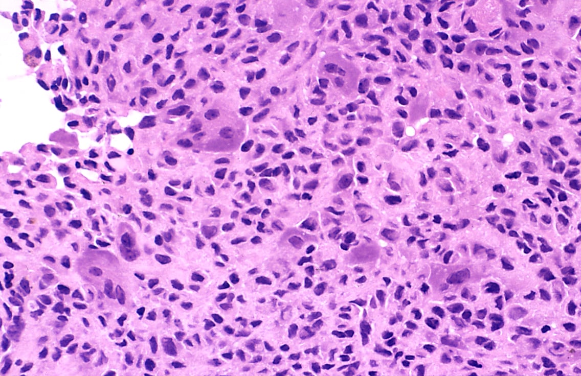
Template:Px Chondroblastoma. H&E stain. | |
|
| |
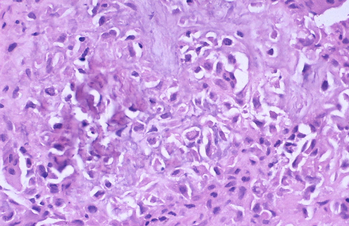
| LM | abundant (chondroid) extracellular material, chondroblasts (variable nuclear morphology (ovoid, folded or grooved), moderate-abundant eosinophilic cytoplasm), +/-calcifications surrounding the cell nests ("chickenwire" appearance) - classic feature, +/-giant cells |
| LM DDx | giant cell tumour of bone, chondroma, well-differentiated chondrosarcoma |
| Site | growth plate - see chondro-osseous tumours |
|
| |
| Clinical history | "young" - growth plates open |
| Symptoms | painful |
| Prognosis | benign |
Chondroblastoma is a benign chondro-osseous tumour that afflicts the young (growth plates open).
General
- Growth plate lesion.
- Sclerotic margin.
- "Young" = growth plates open.
- Most common in teens.
- Typically painful.[1]
- Radiographic osteolytic lesion of the epiphysis [1]
- Rare, 1 % of all bone tumors
- Benign
- Humerus, tibia, femur
Gross
- Well-defined lesion.
Image
Microscopic
Features:[2]
- Abundant extracellular material - pink on H&E stain - looks vaguely like cartilage.
- Sometimes described as 'immature cartilage' (very narrow DDX for this type of cartilage)
- Chondroblasts:
- Nuclear morphology variable: ovoid, folded or grooved.
- Moderate-abundant eosinophilic cytoplasm.
- +/-Calcification surrounds the cell nests ("chickenwire" appearance) - classic feature.
- Cell nests have a thin pale blue rimming.
- +/-Giant cells.
- May lead to confusion with giant cell tumour of bone.
- Not infrequently associated with an aneurysmal bone cyst (33%).[3]
DDx:
- Giant cell tumour of bone.
- Chondroma.
- Well-differentiated chondrosarcoma.
- Chondromyxoid fibroma - also has 'immature cartilage'
- Aneurysmal bone cyst - dont forget that these may be secondary to another lesion.
Images
- Chondroblastoma - intermed mag.jpg
Chondroblastoma - intermed. mag. (WC)
- Chondroblastoma - very high mag.jpg
Chondroblastoma - very high mag. (WC)
- Bone Chondroblastoma Medium Power.jpg
Chicken wire calcification. (SKB)
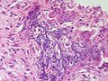
Chicken wire calcification. (SKB)
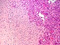
Immature cartilage (left) abutting aneurysmal bone cyst (right) with giant cells. (SKB)
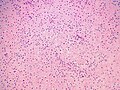
Immature cartilage]]
Immature cartilage surrounded by aneurysmal bone cyst with giant cells. (SKB)
Immature cartilage surrounded by aneurysmal bone cyst with giant cells. (SKB)


www:
- Chondroblastoma (medscape.com).[4]
- Chondroblastoma with "chickenwire" appearance (medscape.com).[4]
- Chondroblastoma (upmc.edu).[5]

IHC
Features:[2]
- S100 +ve.
- Vimentin +ve.[4]
See also
References
- ↑ Mitchell, Richard; Kumar, Vinay; Fausto, Nelson; Abbas, Abul K.; Aster, Jon (2011). Pocket Companion to Robbins & Cotran Pathologic Basis of Disease (8th ed.). Elsevier Saunders. pp. 625. ISBN 978-1416054542.
- ↑ 2.0 2.1 Humphrey, Peter A; Dehner, Louis P; Pfeifer, John D (2008). The Washington Manual of Surgical Pathology (1st ed.). Lippincott Williams & Wilkins. pp. 642. ISBN 978-0781765275.
- ↑ Sepah, YJ.; Umer, M.; Minhas, K.; Hafeez, K. (2007). "Chondroblastoma of the cuboid with an associated aneurysmal bone cyst: a case report.". J Med Case Rep 1: 135. doi:10.1186/1752-1947-1-135. PMID 17999776.
- ↑ 4.0 4.1 4.2 URL: http://emedicine.medscape.com/article/1254949-diagnosis. Accessed on: 31 December 2010.
- ↑ URL: http://path.upmc.edu/cases/case494.html. Accessed on: 24 January 2012.