Difference between revisions of "Gunshot wounds"
Jump to navigation
Jump to search
| (5 intermediate revisions by the same user not shown) | |||
| Line 1: | Line 1: | ||
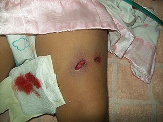
[[Image:Gunshot_wonud_to_leg.JPG|thumb|right|Gunshot wound to the leg showing entrance and exit. (WC)]] | |||
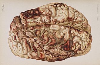
[[Image:Hofmann Lehrbuch brain gunshot.jpg|thumb|right|Depiction of a gunshot wound of the brain. (WC/National Library of Medicine)]] | [[Image:Hofmann Lehrbuch brain gunshot.jpg|thumb|right|Depiction of a gunshot wound of the brain. (WC/National Library of Medicine)]] | ||
This article deals with '''gunshot wounds''', which are seen by (forensic) pathologists in the context of forensic autopsies. | This article deals with '''gunshot wounds''', which are seen by (forensic) pathologists in the context of forensic autopsies. | ||
| Line 4: | Line 5: | ||
An introduction to forensic pathology is in the ''[[forensic pathology]]'' article. | An introduction to forensic pathology is in the ''[[forensic pathology]]'' article. | ||
== | ==Bullet accounting== | ||
Number of entrance wounds should equal the number of exit wounds. | Number of entrance wounds should equal the number of exit wounds. | ||
| Line 26: | Line 27: | ||
==Firearm projectiles== | ==Firearm projectiles== | ||
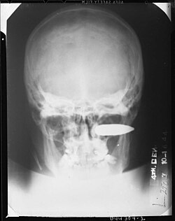
[[Image:260529196 232ebabc57 bBalleIncendiaire.jpg|thumb|200px|X-rays are useful for locating bullets. (WC)]] | |||
===Evidence=== | ===Evidence=== | ||
*Bullets are often good evidence: | *Bullets are often good evidence: | ||
**The calibre (size) and markings from the barrel (on handgun/rifle projectiles) allow it to be matched to the weapon that fired it. | **The calibre (size) and markings from the barrel (on handgun/rifle projectiles) allow it to be matched to the weapon that fired it. | ||
**Thus, all projectiles are recovered from a body... and it is | **Thus, all projectiles are recovered from a body... and it is routine to X-ray all gunshot victims. | ||
===Classification=== | ===Classification=== | ||
| Line 62: | Line 64: | ||
===Images=== | ===Images=== | ||
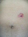
====Abdomen==== | |||
<gallery> | |||
Image:Single_gunshot_abdomen.jpg | Entrance wound - abdomen. (WC) | |||
</gallery> | |||
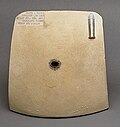
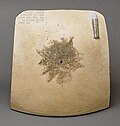
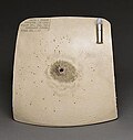
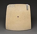
====Ceramic plates (experimental)==== | |||
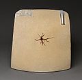
<gallery> | <gallery> | ||
Image: Experimental gunshot entrance wound - dist to target 0 inch.jpg | Exp. GSW, ent. contact (WC) | Image: Experimental gunshot entrance wound - dist to target 0 inch.jpg | Exp. GSW, ent. contact (WC) | ||
| Line 68: | Line 75: | ||
Image: Experimental gunshot entrance wound - dist to target 6 inch.jpg | Exp. GSW, ent. 6" (WC) | Image: Experimental gunshot entrance wound - dist to target 6 inch.jpg | Exp. GSW, ent. 6" (WC) | ||
Image: Experimental gunshot entrance wound - dist to target 25 inch.jpg | Exp. GSW, ent. 25" (WC) | Image: Experimental gunshot entrance wound - dist to target 25 inch.jpg | Exp. GSW, ent. 25" (WC) | ||
</gallery> | </gallery> | ||
| Line 81: | Line 85: | ||
*NO abrasion at wound margin - '''key feature'''. | *NO abrasion at wound margin - '''key feature'''. | ||
*In skull -- outer table defect typically larger than inner table defect (external beveling). | *In skull -- outer table defect typically larger than inner table defect (external beveling). | ||
===Atypical exit wounds=== | ===Atypical exit wounds=== | ||
| Line 92: | Line 91: | ||
*Supporting surface may lead to abrasion. | *Supporting surface may lead to abrasion. | ||
*May appear to be an entrance wound. | *May appear to be an entrance wound. | ||
===Images=== | |||
<gallery> | |||
Image:Gunshot_skull.jpg|Exit wound. (WC) | |||
Image:Experimental gunshot exit wound.jpg |Experimental gunshot exit wound. (WC) | |||
</gallery> | |||
==Special entrance/exit wounds== | ==Special entrance/exit wounds== | ||
Latest revision as of 14:03, 5 January 2016
This article deals with gunshot wounds, which are seen by (forensic) pathologists in the context of forensic autopsies.
An introduction to forensic pathology is in the forensic pathology article.
Bullet accounting
Number of entrance wounds should equal the number of exit wounds.
If the above is not so... the explanation is:
- Bullets are still in the body.
- "Tandem bullets" -- two bullets entered (or exited) at the same place.
- Secondary projectile -- a bullet hit something, e.g. bone, and made it fly out of the body.
- Pathologist missed an entrance or exit.
- Places to look:
- Below chin (common in suicides).
- In the mouth (common in suicides).
- Ears.
- Back.
- Places to look:
"Sites of election" (suicide)
Common places where people shoot themselves:[1]
- Mouth.
- Temple.
- Left chest.
- Below the chin.
Firearm projectiles
Evidence
- Bullets are often good evidence:
- The calibre (size) and markings from the barrel (on handgun/rifle projectiles) allow it to be matched to the weapon that fired it.
- Thus, all projectiles are recovered from a body... and it is routine to X-ray all gunshot victims.
Classification
Two broad groups:
- Shotgun projectiles.[2]
- Buckshot - usu. 7-9 pellets, typically 6-9 mm (diameter).
- Birdshot - many pellets, typically 2-5 mm (diameter).
- Slug - one large bullet; may be confused with a (high power) rifle projectile.
- Handgun/rifle projectiles.
- Similar in size to the barrel - large when compared to shotgun projectiles.
- Bullets from handguns/rifles are marked by the barrel on the way-out (by grooves which in part spin on it to improve accuracy).
Subclassification
- Rifled projectile are often group by diameter, e.g. "22 caliber" is 0.22 inches (~5.6 mm).
Entrance wounds
Characteristics:[3]
- Circular/round defect --especially if the projectile strikes at a right angle to the surface.
- If the projectile strikes at an angle the injury will be elliptical and the long axis of the ellipse will lie approximately in the plane the bullet traveled.
- An abrasion, or scraping, --concentric or eccentric-- usually surrounds a deep wound (key feature -- used to differentiate from exit wounds).
- Eccentric abrasion suggest directionality.
- Usually smaller than exit wounds.
- In skull the inner table defect is typically larger than outer table defect ("internal bevel").
Atypical entrance wounds
- Irregular (non-circular/non-elliptical) margin.
- May be due to close range/contact.
- Classically results in a "stellate" pattern.
- Bullet ricochets --hits other object before hitting target, gun defective -- bullet's long axis doesn't coincide with its velocity vector.
- Classically results in a "D-shaped" wound.[3]
- May be due to close range/contact.
Images
Abdomen
Ceramic plates (experimental)
Exit wounds
Characteristics:
- Usually bigger than entrance wounds.
- Morphologic shape -- variable/irregular.
- May be : stellate, ovoid, round.
- Looks like it may be easily approximated with sutures -- unlike a typical entrance wound.
- NO abrasion at wound margin - key feature.
- In skull -- outer table defect typically larger than inner table defect (external beveling).
Atypical exit wounds
"Shored" exit wounds:
- Exit defect created whilst surface supported/adjacent to firm surface.
- Supporting surface may lead to abrasion.
- May appear to be an entrance wound.
Images
Special entrance/exit wounds
Keyed wounds:
- Combination entrance/exit wounds -- result from a bullet grazing the victim.
- Attached skin tags can give directionality;[4] they are classically found at the aspect of the wound distant from the projectile's origin.
- The University of Utah describes it as:[5]
- Characteristically, the side of the [skin] tag demonstrating a laceration is the side of the projection toward the weapon.
Distance of shooter
Contact
- Muzzle impression.
- Stellate splitting/tearing of the skin -- especially if it overlies a bony surface.
- Soot/gun powder residue - deep in the wound.
Close range
- Stippling (tattooing) - punctate abrasions around the entrance wound.[6]
- Suggests a distance > 1 cm and <= 60 cm (rim fire cartridge), > 1 cm and <= 90-120 cm (centrefire cartridge).[7]
- Soot/gun powder residue - dirt at the entrance, can be wiped-off.
- Scalloped border/edge in shotgun wound.
Intermediate
- Stippling.
- No soot.
Shotgun:
- "Satellite" wounds.
- Wad marks - typically have sharp edges +/- sharp corners.
Indeterminate/Distant
- Previously known as distant for non-shotguns, as this is the pattern seen if a shooter is far from the victim.
- No soot.
- No stippling.
Notes:
- Absence of soot & stippling does not exclude near range; the lack of soot and stippling may be explained by clothing or an intermediate target that the projectile travelled through prior to striking the victim.
- Intermediate should not be confused with indeterminate.
Summary tables
Summary table - hand gun entry wounds:
| Distance | Skin splitting | Soot | Stippling | Other |
|---|---|---|---|---|
| Contact | +/- (only if to head) | + (in wound) | + (in wound) | muzzle imprint |
| Near contact | - | + (in & around wound) | + | no muzzle imprint |
| Intermediate | - | - | + | should not be confused with intermediate target |
| Indeterminate | - | - | - | previous known as distant |
Summary table - shot gun entry wounds:
| Distance | Skin splitting | Soot | Stippling | Projectile topography | Wad mark |
|---|---|---|---|---|---|
| Contact | +/- (only if to head) | + (in wound) | + (in wound) | round | - |
| Near contact | - | + (in & around wound | + (in & around wound) | round with scalloped border | - |
| Intermediate | - | - | + | round with scalloped border + "satellite" bullets | +/- |
| Distant | - | - | - | scattered, multiple round-same size | - |
Injury severity due to GSWs
The damage of a projectile depends on:
- Where the bullet strikes, e.g. ascending aorta vs. brain vs. tibia vs. gluteus maximus.
- Kinetic energy of the bullet.
-
- Velocity is more important -- as it is squared (duh).
-
- Cavitation effect; temporary cavity formation.[8]
See also
References
- ↑ Rouse D, Dunn L (September 1992). "Firearm fatalities". Forensic Sci. Int. 56 (1): 59–64. PMID 1398378.
- ↑ DiMaio, Vincent J.M.; Dana, Suzanna E. (2006). Handbook of Forensic Pathology (2nd ed.). CRC Press. pp. 147. ISBN 978-0849392870.
- ↑ 3.0 3.1 Denton JS, Segovia A, Filkins JA (September 2006). "Practical pathology of gunshot wounds". Arch. Pathol. Lab. Med. 130 (9): 1283?9. PMID 16948512. http://journals.allenpress.com/jrnlserv/?request=get-abstract&issn=0003-9985&volume=130&page=1283.
- ↑ Dixon, DS. (Jan 1984). "Determination of direction of fire from graze gunshot wounds of internal organs.". J Forensic Sci 29 (1): 331-5. PMID 6699600.
- ↑ URL: http://library.med.utah.edu/WebPath/TUTORIAL/GUNS/GUNINJ.html. Accessed on: 6 March 2012.
- ↑ DiMaio, Vincent J.M.; Dana, Suzanna E. (2006). Handbook of Forensic Pathology (2nd ed.). CRC Press. pp. 135. ISBN 978-0849392870.
- ↑ DiMaio, Vincent J.M.; Dana, Suzanna E. (2006). Handbook of Forensic Pathology (2nd ed.). CRC Press. pp. 139. ISBN 978-0849392870.
- ↑ Maiden N (2009). "Ballistics reviews: mechanisms of bullet wound trauma". Forensic Sci Med Pathol 5 (3): 204–9. doi:10.1007/s12024-009-9096-6. PMID 19644779.
