Difference between revisions of "Chordoma"
Jump to navigation
Jump to search
(wiify) |
|||
| (21 intermediate revisions by 3 users not shown) | |||
| Line 1: | Line 1: | ||
{{ Infobox diagnosis | {{ Infobox diagnosis | ||
| Name = {{PAGENAME}} | | Name = {{PAGENAME}} | ||
| Image = Chordoma_-_high_mag.jpg | | Image = Chordoma_-_high_mag.jpg | ||
| Width = | | Width = | ||
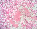
| Caption = Chordoma. [[HPS stain]]. | | Caption = Chordoma. [[HPS stain]]. | ||
| Micro = ''physaliphorous cells'' (also ''bubble cells'') - very large clear bubble with a sharp border, bubble does not compress nucleus; islands of cells surrounded by fibrous tissue; myxoid background | | Micro = ''physaliphorous cells'' (also ''bubble cells'') - very large clear bubble with a sharp border, bubble does not compress nucleus; islands of cells surrounded by fibrous tissue; myxoid background | ||
| Subtypes = | | Subtypes = | ||
| LMDDx = [[chondrosarcoma]], [[myxoid lesions]], [[parachordoma]] | | LMDDx = [[chondrosarcoma]], [[myxoid lesions]], [[parachordoma]], chordoid lesions (e.g. chordoid glioma, [[chordoid meningioma]]), [[metastasis]] (e.g. [[clear cell renal cell carcinoma]]) | ||
| Stains = | | Stains = | ||
| IHC = S-100 +ve, AE1/AE3 +ve, Brachyury +ve, EMA +ve | | IHC = S-100 +ve, [[AE1/AE3]] +ve, Brachyury +ve, [[EMA]] +ve | ||
| EM = | | EM = | ||
| Molecular = | | Molecular = | ||
| Line 32: | Line 32: | ||
==General== | ==General== | ||
*Location: usually sacrum or clivus. | *Location: usually sacrum or clivus. | ||
*It is a [[bone tumour]]. | *It is a malignant [[bone tumour]] (1-4% of all primary bone tumors). | ||
*Usually after age 30. | |||
===Classification=== | |||
* Chordoma, NOS (ICD-O: 9370/3). | |||
* Chondroid chordoma (ICD-O: 9371/3). | |||
* Dedifferentiated chordoma (ICD-O: 9372/3). | |||
==Gross== | ==Gross== | ||
| Line 54: | Line 60: | ||
DDx: | DDx: | ||
*[[Chondrosarcoma]]. | *[[Chondrosarcoma]] - negative for EMA and cytokeratins. Beware of 'chondroid' chordoma. | ||
*[[Myxoid lesions]]. | *[[Myxoid lesions]]. | ||
**Myxopapillary ependymoma. | **[[Myxopapillary ependymoma]]. | ||
**Myxoid liposarcoma | **[[Myxoid liposarcoma]] - negative for EMA and cytokeratins. | ||
* | *Chordoid lesions. | ||
** | **[[Chordoid meningioma]]. | ||
** | **Chordoid glioma<ref>{{Cite journal | last1 = Zarghouni | first1 = M. | last2 = Vandergriff | first2 = C. | last3 = Layton | first3 = KF. | last4 = McGowan | first4 = JB. | last5 = Coimbra | first5 = C. | last6 = Bhakti | first6 = A. | last7 = Opatowsky | first7 = MJ. | title = Chordoid glioma of the third ventricle. | journal = Proc (Bayl Univ Med Cent) | volume = 25 | issue = 3 | pages = 285-6 | month = Jul | year = 2012 | doi = | PMID = 22754136 }}</ref> - location, location, location. | ||
**Large notochordal rest. | **Large notochordal rest - only evidence of destructive growth can identify a chordoma. | ||
*Metastasis | *[[Metastasis]]. | ||
**Metastatic signet ring cell | **Metastatic [[signet ring cell carcinoma]] - negative for S100 and brachyury, [[clinical history]] (important). | ||
**Metastatic clear cell renal cell carcinoma. | **Metastatic [[clear cell renal cell carcinoma]] - negative for S100 and brachyury, clinical history (important). | ||
*[[Parachordoma]] - extremely rare. | *[[Parachordoma]] - extremely rare. | ||
===Images=== | ===Images=== | ||
====Case 1==== | |||
<gallery> | <gallery> | ||
Image:Chordoma_-_low_mag.jpg | Chordoma - low mag. (WC) | Image:Chordoma_-_low_mag.jpg | Chordoma - low mag. (WC) | ||
| Line 73: | Line 80: | ||
Image:Chordoma_-_high_mag.jpg | Chordoma - high mag. (WC) | Image:Chordoma_-_high_mag.jpg | Chordoma - high mag. (WC) | ||
Image:Chordoma - very high mag.jpg| Chordoma - very high mag. (WC) | Image:Chordoma - very high mag.jpg| Chordoma - very high mag. (WC) | ||
</gallery> | |||
====Case 2==== | |||
<gallery> | |||
Image: Chordoma -- low mag.jpg | Chordoma - low mag. (WC) | |||
Image: Chordoma -- intermed mag.jpg | Chordoma - intermed. mag. (WC) | |||
Image: Chordoma - alt -- intermed mag.jpg | Chordoma - intermed. mag. (WC) | |||
Image: Chordoma -- high mag.jpg | Chordoma - high mag. (WC) | |||
</gallery> | |||
====Case 3==== | |||
<gallery> | |||
Image:Bone Chordoma 2 HP2.JPG|Physaliphorous cells. (SKB) | Image:Bone Chordoma 2 HP2.JPG|Physaliphorous cells. (SKB) | ||
Image:Bone Chordoma 2 LP PA.JPG|Low power view. Somewhat lobulated tumour with loose areas, cellular areas and fibrous septa. (SKB) | Image:Bone Chordoma 2 LP PA.JPG|Low power view. Somewhat lobulated tumour with loose areas, cellular areas and fibrous septa. (SKB) | ||
| Line 80: | Line 97: | ||
Image:Bone Chordoma HP PA.JPG|Chordoma cells may form sheets of cells with eosinophilic cytoplasm. (SKB) | Image:Bone Chordoma HP PA.JPG|Chordoma cells may form sheets of cells with eosinophilic cytoplasm. (SKB) | ||
</gallery> | </gallery> | ||
www | ====www==== | ||
*[http://path.upmc.edu/cases/case64.html Chordoma (upmc.edu)]. | *[http://path.upmc.edu/cases/case64.html Chordoma (upmc.edu)]. | ||
*[http://path.upmc.edu/cases/case312/micro.html Chordoma - sacrum - several images (upmc.edu)]. | *[http://path.upmc.edu/cases/case312/micro.html Chordoma - sacrum - several images (upmc.edu)]. | ||
| Line 87: | Line 104: | ||
==IHC== | ==IHC== | ||
Features:<ref>URL: [http://path.upmc.edu/cases/case312/micro.html http://path.upmc.edu/cases/case312/micro.html]. Accessed on: 14 January 2012.</ref><ref name=pmid2431128>{{Cite journal | last1 = Coindre | first1 = JM. | last2 = Rivel | first2 = J. | last3 = Trojani | first3 = M. | last4 = De Mascarel | first4 = I. | last5 = De Mascarel | first5 = A. | title = Immunohistological study in chordomas. | journal = J Pathol | volume = 150 | issue = 1 | pages = 61-3 | month = Sep | year = 1986 | doi = 10.1002/path.1711500110 | PMID = 2431128 }}</ref> | Features:<ref>URL: [http://path.upmc.edu/cases/case312/micro.html http://path.upmc.edu/cases/case312/micro.html]. Accessed on: 14 January 2012.</ref><ref name=pmid2431128>{{Cite journal | last1 = Coindre | first1 = JM. | last2 = Rivel | first2 = J. | last3 = Trojani | first3 = M. | last4 = De Mascarel | first4 = I. | last5 = De Mascarel | first5 = A. | title = Immunohistological study in chordomas. | journal = J Pathol | volume = 150 | issue = 1 | pages = 61-3 | month = Sep | year = 1986 | doi = 10.1002/path.1711500110 | PMID = 2431128 }}</ref> | ||
*S-100 +ve. | *S-100 +ve - '''important'''. | ||
*AE1/AE3 +ve. | *[[AE1/AE3]] +ve - '''important'''. | ||
*Brachyury +ve | *Brachyury +ve - '''key stain'''. | ||
**Protein important for axial development, affects notochord development.<ref name=omim601397>{{OMIM|601397}}</ref> | **Protein important for axial development, affects notochord development.<ref name=omim601397>{{OMIM|601397}}</ref> | ||
**''Brachyury'' literally means ''short tail''.<ref>URL: [http://www.jstor.org/pss/86845 http://www.jstor.org/pss/86845]. Accessed on: 18 May 2010.</ref> | **''Brachyury'' literally means ''short tail''.<ref>URL: [http://www.jstor.org/pss/86845 http://www.jstor.org/pss/86845]. Accessed on: 18 May 2010.</ref> | ||
*EMA +ve. | *[[EMA]] +ve. | ||
*[[PAX8]] -ve (1 +ve (weak) of 12.<ref name=pmid21102418>{{Cite journal | last1 = Sangoi | first1 = AR. | last2 = Karamchandani | first2 = J. | last3 = Lane | first3 = B. | last4 = Higgins | first4 = JP. | last5 = Rouse | first5 = RV. | last6 = Brooks | first6 = JD. | last7 = McKenney | first7 = JK. | title = Specificity of brachyury in the distinction of chordoma from clear cell renal cell carcinoma and germ cell tumors: a study of 305 cases. | journal = Mod Pathol | volume = 24 | issue = 3 | pages = 425-9 | month = Mar | year = 2011 | doi = 10.1038/modpathol.2010.196 | PMID = 21102418 }}</ref>). | |||
*Other keratins:<ref name=pmid9195570>{{Cite journal | last1 = Naka | first1 = T. | last2 = Iwamoto | first2 = Y. | last3 = Shinohara | first3 = N. | last4 = Chuman | first4 = H. | last5 = Fukui | first5 = M. | last6 = Tsuneyoshi | first6 = M. | title = Cytokeratin subtyping in chordomas and the fetal notochord: an immunohistochemical analysis of aberrant expression. | journal = Mod Pathol | volume = 10 | issue = 6 | pages = 545-51 | month = Jun | year = 1997 | doi = | PMID = 9195570 }}</ref> | |||
**CK8 +ve (16/16). | |||
**CK19 +ve (16/16). | |||
**CK18 +ve/-ve (9 +ve/16). | |||
**[[CK7]] -ve (1 +ve/16). | |||
**[[CK20]] -ve (0 +ve/16). | |||
Classic chordoma panel:<ref name=pmid24296480>{{Cite journal | last1 = Shen | first1 = J. | last2 = Shi | first2 = Q. | last3 = Lu | first3 = J. | last4 = Wang | first4 = DL. | last5 = Zou | first5 = TM. | last6 = Yang | first6 = HL. | last7 = Zhu | first7 = GQ. | title = Histological study of chordoma origin from fetal notochordal cell rests. | journal = Spine (Phila Pa 1976) | volume = 38 | issue = 25 | pages = 2165-70 | month = Dec | year = 2013 | doi = 10.1097/BRS.0000000000000010 | PMID = 24296480 }}</ref> | |||
*EMA, AE1/AE3, CAM5.2, vimentin, S-100. | |||
Key points: | |||
*Brachyury is not a commonly stocked antibody. | |||
*Chordoma will be S100 '''''and''''' epithelial marker positive. | |||
*Many other items in the DDx will be either S100 '''''or''''' epithelial marker positive. | |||
==See also== | ==See also== | ||
*[[CNS tumours]]. | *[[CNS tumours]]. | ||
*[[Parachordoma]]. | |||
==References== | ==References== | ||
Latest revision as of 19:36, 31 July 2016
| Chordoma | |
|---|---|
| Diagnosis in short | |
 Chordoma. HPS stain. | |
|
| |
| LM | physaliphorous cells (also bubble cells) - very large clear bubble with a sharp border, bubble does not compress nucleus; islands of cells surrounded by fibrous tissue; myxoid background |
| LM DDx | chondrosarcoma, myxoid lesions, parachordoma, chordoid lesions (e.g. chordoid glioma, chordoid meningioma), metastasis (e.g. clear cell renal cell carcinoma) |
| IHC | S-100 +ve, AE1/AE3 +ve, Brachyury +ve, EMA +ve |
| Gross | myxoid |
| Site | sacrum or clivus |
|
| |
| Prevalence | uncommon |
Chordoma is an uncommon tumour in neuropathology.
General
- Location: usually sacrum or clivus.
- It is a malignant bone tumour (1-4% of all primary bone tumors).
- Usually after age 30.
Classification
- Chordoma, NOS (ICD-O: 9370/3).
- Chondroid chordoma (ICD-O: 9371/3).
- Dedifferentiated chordoma (ICD-O: 9372/3).
Gross
- Soft, gelatinous, lobulated.[1]
DDx:
- Bony metastasis (mucinous carcinoma) - typically multifocal.
Image:
Microscopic
Features:[2]
- Architecture: islands of cells surrounded by fibrous tissue.
- Also described as "lobulated" architecture; may not be apparent.
- Myxoid background - grey extracellular material, variable amount present.
- Mixed cell population:
- Abundant eosinophilic cytoplasm.
- Physaliphorous cells or bubble cells - key feature.
- Have a very large clear bubble with a sharp border; bubble does not compress nucleus - nucleus may be in bubble.
DDx:
- Chondrosarcoma - negative for EMA and cytokeratins. Beware of 'chondroid' chordoma.
- Myxoid lesions.
- Myxopapillary ependymoma.
- Myxoid liposarcoma - negative for EMA and cytokeratins.
- Chordoid lesions.
- Chordoid meningioma.
- Chordoid glioma[3] - location, location, location.
- Large notochordal rest - only evidence of destructive growth can identify a chordoma.
- Metastasis.
- Metastatic signet ring cell carcinoma - negative for S100 and brachyury, clinical history (important).
- Metastatic clear cell renal cell carcinoma - negative for S100 and brachyury, clinical history (important).
- Parachordoma - extremely rare.
Images
Case 1
Chordoma - low mag. (WC)
Chordoma - intermed. mag. (WC)
Chordoma - high mag. (WC)
Chordoma - very high mag. (WC)
Case 2
Chordoma - low mag. (WC)
Chordoma - intermed. mag. (WC)
Chordoma - intermed. mag. (WC)
Chordoma - high mag. (WC)
Case 3
Physaliphorous cells. (SKB)
Low power view. Somewhat lobulated tumour with loose areas, cellular areas and fibrous septa. (SKB)
Physaliphorous cells. (SKB)
Physaliphorous cells. (SKB)
Physaliphorous cells.(SKB)
Chordoma cells may form sheets of cells with eosinophilic cytoplasm. (SKB)

www
- Chordoma (upmc.edu).
- Chordoma - sacrum - several images (upmc.edu).
- Chordoma - additional case with several images (upmc.edu).
IHC
- S-100 +ve - important.
- AE1/AE3 +ve - important.
- Brachyury +ve - key stain.
- EMA +ve.
- PAX8 -ve (1 +ve (weak) of 12.[8]).
- Other keratins:[9]
Classic chordoma panel:[10]
- EMA, AE1/AE3, CAM5.2, vimentin, S-100.
Key points:
- Brachyury is not a commonly stocked antibody.
- Chordoma will be S100 and epithelial marker positive.
- Many other items in the DDx will be either S100 or epithelial marker positive.
See also
References
- ↑ URL: http://www.histopathology-india.net/Chordoma.htm. Accessed on: 12 April 2012.
- ↑ Tadrous, Paul.J. Diagnostic Criteria Handbook in Histopathology: A Surgical Pathology Vade Mecum (1st ed.). Wiley. pp. 184. ISBN 978-0470519035.
- ↑ Zarghouni, M.; Vandergriff, C.; Layton, KF.; McGowan, JB.; Coimbra, C.; Bhakti, A.; Opatowsky, MJ. (Jul 2012). "Chordoid glioma of the third ventricle.". Proc (Bayl Univ Med Cent) 25 (3): 285-6. PMID 22754136.
- ↑ URL: http://path.upmc.edu/cases/case312/micro.html. Accessed on: 14 January 2012.
- ↑ Coindre, JM.; Rivel, J.; Trojani, M.; De Mascarel, I.; De Mascarel, A. (Sep 1986). "Immunohistological study in chordomas.". J Pathol 150 (1): 61-3. doi:10.1002/path.1711500110. PMID 2431128.
- ↑ Online 'Mendelian Inheritance in Man' (OMIM) 601397
- ↑ URL: http://www.jstor.org/pss/86845. Accessed on: 18 May 2010.
- ↑ Sangoi, AR.; Karamchandani, J.; Lane, B.; Higgins, JP.; Rouse, RV.; Brooks, JD.; McKenney, JK. (Mar 2011). "Specificity of brachyury in the distinction of chordoma from clear cell renal cell carcinoma and germ cell tumors: a study of 305 cases.". Mod Pathol 24 (3): 425-9. doi:10.1038/modpathol.2010.196. PMID 21102418.
- ↑ Naka, T.; Iwamoto, Y.; Shinohara, N.; Chuman, H.; Fukui, M.; Tsuneyoshi, M. (Jun 1997). "Cytokeratin subtyping in chordomas and the fetal notochord: an immunohistochemical analysis of aberrant expression.". Mod Pathol 10 (6): 545-51. PMID 9195570.
- ↑ Shen, J.; Shi, Q.; Lu, J.; Wang, DL.; Zou, TM.; Yang, HL.; Zhu, GQ. (Dec 2013). "Histological study of chordoma origin from fetal notochordal cell rests.". Spine (Phila Pa 1976) 38 (25): 2165-70. doi:10.1097/BRS.0000000000000010. PMID 24296480.