Difference between revisions of "Parathyroid carcinoma"
Jump to navigation
Jump to search
| Line 1: | Line 1: | ||
{{ Infobox diagnosis | {{ Infobox diagnosis | ||
| Name = {{PAGENAME}} | | Name = {{PAGENAME}} | ||
| Image = | | Image = Parathyroid carcinoma - 1 -- high mag.jpg | ||
| Width = | | Width = | ||
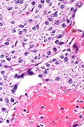
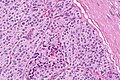
| Caption = | | Caption = Parathyroid carcinoma. [[H&E stain]]. | ||
| Synonyms = | | Synonyms = | ||
| Micro = | | Micro = | ||
Revision as of 04:02, 11 June 2019
| Parathyroid carcinoma | |
|---|---|
| Diagnosis in short | |
 Parathyroid carcinoma. H&E stain. | |
| IHC | PAX8 +ve, Ki-67 >6% +ve |
| Site | parathyroid gland |
|
| |
| Prevalence | very rare |
| Prognosis | poor |
| Clin. DDx | parathyroid adenoma, parathyroid hyperplasia, thyroid cancer |
| Treatment | surgical excision |
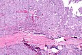
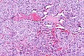
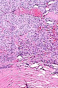
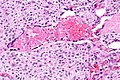
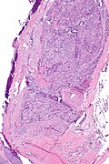
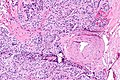
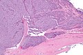
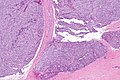
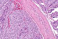
Parathyroid carcinoma is a rare epithelial malignancy of the parathyroid gland.
General
- Extremely rare.
Microscopic
Features:[1]
- Histologically normal parathyroid cells.
- Cytologic features not reliable for diagnosis.
- Fibrous capsule.
- Invasion of surrounding tissue - key feature.
- +/-Metastasis - diagnostic feature.
Note:
- Diagnosis of parathyroid carcinoma is like that of malignant pheochromocytoma - cytology useless, tissue invasion and metastases are the key features.
Images
PC - low mag. (WC)
PC - intermed. mag. (WC)
PC - intermed. mag. (WC)
PC - high mag. (WC)
PC - very high mag. (WC)
PC - low mag. (WC)
PC - intermed. mag. (WC)
PC - very low mag. (WC)
PC - low mag. (WC)
PC - intermed. mag. (WC)
PC - high mag. (WC)


IHC
- Ki-67 >6% of cells positive - supports diagnosis.[2]
- Parathyroid adenomas and hyperplasias ~ 3%.
- PAX8 +ve.[3]
See also
References
- ↑ Kumar, Vinay; Abbas, Abul K.; Fausto, Nelson; Aster, Jon (2009). Robbins and Cotran pathologic basis of disease (8th ed.). Elsevier Saunders. pp. 1128. ISBN 978-1416031215.
- ↑ Abbona, GC.; Papotti, M.; Gasparri, G.; Bussolati, G. (Feb 1995). "Proliferative activity in parathyroid tumors as detected by Ki-67 immunostaining.". Hum Pathol 26 (2): 135-8. PMID 7860042.
- ↑ Ordóñez, NG. (May 2012). "Value of PAX 8 immunostaining in tumor diagnosis: a review and update.". Adv Anat Pathol 19 (3): 140-51. doi:10.1097/PAP.0b013e318253465d. PMID 22498579.