Difference between revisions of "Histology artifacts"
Jump to navigation
Jump to search
(→Coverslip artifact: +images) |
|||
| Line 52: | Line 52: | ||
===Images=== | ===Images=== | ||
====Case 1==== | |||
<gallery> | <gallery> | ||
Image: Epididymis with coverslip artifact -- low mag.jpg | CSA - low mag. (WC) | Image: Epididymis with coverslip artifact -- low mag.jpg | CSA - low mag. (WC) | ||
| Line 58: | Line 59: | ||
Image: Epididymis with coverslip artifact - wc -- high mag.jpg | CSA - high mag. (WC) | Image: Epididymis with coverslip artifact - wc -- high mag.jpg | CSA - high mag. (WC) | ||
Image: Epididymis with coverslip artifact - wc -- very high mag.jpg | CSA - very high mag. (WC) | Image: Epididymis with coverslip artifact - wc -- very high mag.jpg | CSA - very high mag. (WC) | ||
</gallery> | |||
====Case 2==== | |||
<gallery> | |||
Image: Coverslip artifact -- intermed mag.jpg | CA - intermed. mag. (WC) | |||
Image: Coverslip artifact -- high mag.jpg | CA - high mag. (WC) | |||
Image: Coverslip artifact - alt -- high mag.jpg | CA - high mag. (WC) | |||
</gallery> | </gallery> | ||
Revision as of 02:35, 1 January 2020
Histology artifacts are common.
Cautery artifact
Main article: Cautery artifact
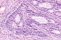
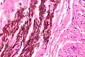
Tissue fold
- Darker well demarcated line with apparent disruption of the architecture.
Images
Tissue fold (center of image) in basal cell hyperplasia of the prostate.
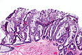
Extensive tissue folding in a SSA.


Chatter artifact
- Fine parallel lines.
Images
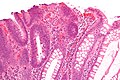
Chatter artifact in tubular adenoma.
Chatter artifact in complex endometrial hyperplasia.
Chatter artifact in TILs.


Tissue tearing
- Usually due to something intrinsic to the tissue that is hard, e.g. calcium.
Image
Tearing due to a calcification.

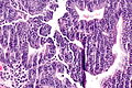
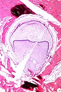
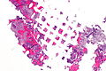
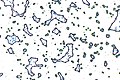
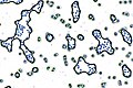
Sponge artifact
- Angulated pieces of tissue with punched-out missing fragments.[1]
- May result in little squares of tissue arranged in a regular pattern.
Images
Sponge artifact - very low mag.
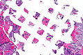
Sponge artifact - low mag.
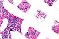
Sponge artifact - intermed. mag.


Bubble artifact
Large
- Large bubbles of air under the cover slip.
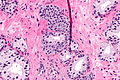
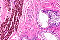
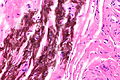
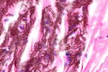
Coverslip artifact
Features:
- Black-grey lines - takes on the outline of the tissue beneath it.
- Above the plane-of-focus of the tissue - key feature.
Images
Case 1
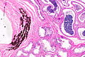
CSA - low mag. (WC)
CSA - intermed mag. (WC)
CSA - high mag. (WC)
CSA - high mag. (WC)
CSA - very high mag. (WC)


Case 2
CA - intermed. mag. (WC)
CA - high mag. (WC)
CA - high mag. (WC)


