Hemangioblastoma
Jump to navigation
Jump to search
| Hemangioblastoma | |
|---|---|
| Diagnosis in short | |
 Cerebellar hemangioblastoma. | |
|
| |
| LM | vascular tumour with large polygonal stromal cells with hyperchromatic nuclei and vacuolar cytoplasm |
| LM DDx | metastatic cell cell renal cell carcinoma |
| IHC | alpha-inhibin +ve, NSE +ve, EMA -ve |
| Site | brain - usu. cerebellum |
|
| |
| Syndromes | von Hippel-Lindau disease |
|
| |
| Prognosis | good (WHO grade I) |
Hemangioblastoma is a low grade brain tumour tumour typically found the cerebellum.
General
- Usually cerebellar.
- Associated with von Hippel-Lindau syndrome.
- WHO grade I.[1]
Microscopic
Features:[2]
- Vascular.
- Polygonal stromal cells with:
- Hyperchromatic nuclei.
- Vacuolar cytoplasm.
DDx:
- Metastatic clear cell renal cell carcinoma.
Images
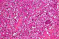
Hemangioblastoma - intermed. mag. (WC)
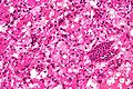
Hemangioblastoma - high mag. (WC)
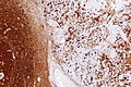
Hemangioblastoma - NSE - intermed. mag. (WC)


www:
IHC
Features:[3]
- Alpha-inhibin +ve (cytoplasm).
- EMA -ve.
- RCC typically +ve.
- NSE +ve (nucleus + cytoplasm).
- RCC typically -ve.
See also
References
- ↑ URL: http://www.expertconsultbook.com/expertconsult/ob/book.do?method=display&type=bookPage&decorator=none&eid=4-u1.0-B978-1-4160-4580-9..00019-8--sc0155&isbn=978-1-4160-4580-9. Accessed on: 9 December 2010.
- ↑ URL: http://emedicine.medscape.com/article/340994-media. Accessed on: 23 June 2010.
- ↑ URL: http://www.nature.com/modpathol/journal/v18/n6/full/3800351a.html. Accessed on: 9 December 2010.